OSMOREGULATION
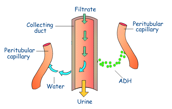
The kidneys play the main role in controlling the water content of our bodies, this is called osmoregulation. Osmoreceptor cells in the hypothalamus monitor the water content of the body. The cell bodies of these nerve cells produce an antidiuretic hormone called ADH. This hormone passes along the axons of the nerve cell leading to the posterior pituitary gland. The amount of ADH released controls how much water is released in urine, if the body isn’t requiring too much water less ADH is released so the water isn’t able to move out of the duct and into the tissue fluid but instead has to be expelled in the urine. If the body does require more water, more ADH is released to make the membrane more water permeable to enable the water to be released into the tissue fluid and not expelled in urine. This means the body retains as much water as it needs and so homeostasis is at work in this process (Jones, Mary and Geoff 1997).
(Andrew biology, 2011)
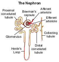
We have two kidneys, each is made up of about a million small filtering units called nephrons. It is in these structures that urine is produced. The starting point is a Bowman’s capsule, a small cup-shaped structure situated in the cortex. The cortex leads to a narrow tube which makes a loop in the medulla back up to the cortex and then joins a collecting duct. The collecting duct transports urine to the pelvis of the kidney where it leaves via the urethra. The nephrons are surrounded by a dense network of capillaries (Brotherton, Judith and Mudie, Kate 1992).
(Caribbeanedu.com, 2010)
The role of the kidney in homeostasis
(Jones, Mary and Geoff 1997).
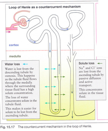
When the blood in the glomerulus of the Bowman’s capsule is filtered, the blood pressure is so high that some of the liquid (the plasma but no blood cells or blood proteins) is forced through the walls of the blood vessels and into the Bowman’s capsule. This plasma contains water, nitrogenous waste e.g. urea, and food materials including glucose, amino acids, vitamins and mineral ions. This liquid is called the glomerular filtrate which then passes down the nephron tubule to the renal medulla where water is released and flows out. As it does this the osmolarity in the tubular is going to increase to make the concentration go from 300 to 1200 milliosmoles. Then as the liquid goes up the right side of the Loop of Henle salt instead water is lost, which makes the concentration 300 milliosmoles again. As it goes through the collecting duct there will either be a high or a low level of ADH do deside if the water is expelled in urine or moves from the duct to the tissue fluid (Marcom Projects Pty, 2008).
The formation of urea in the liver
Urea is a small organic molecule containing nitrogen, is made in the liver from here it passes into the blood. In large concentrations in the blood urea can damage cells and so must be constantly removed. Then as the blood passes into the kidneys the urea is removed from it and excreted from the body in the urine. The liver produces urea from amino acids, the human body is unable to store proteins and amino acids which contain nitrogen and so any excess must be disposed of. Though as amino acids contain useful energy they are converted to carbohydrates or fats which are stored and can also be used in respiration. So the process of converting amino acids to carbohydrates or fat is called deamination and this takes place in the liver. Nitrogen is removed from the amino acid thus producing ammonia, this being an extremely soluble and very toxic substance capable of damaging cells, cannot be allowed to accumulate. It is therefore converted to urea by combining it with carbon dioxide (Brotherton, Judith and Mudie, Kate 1992).

